Chest Drain & Needle Decompression Trainer with Augmented Reality Training (ART)
02:04
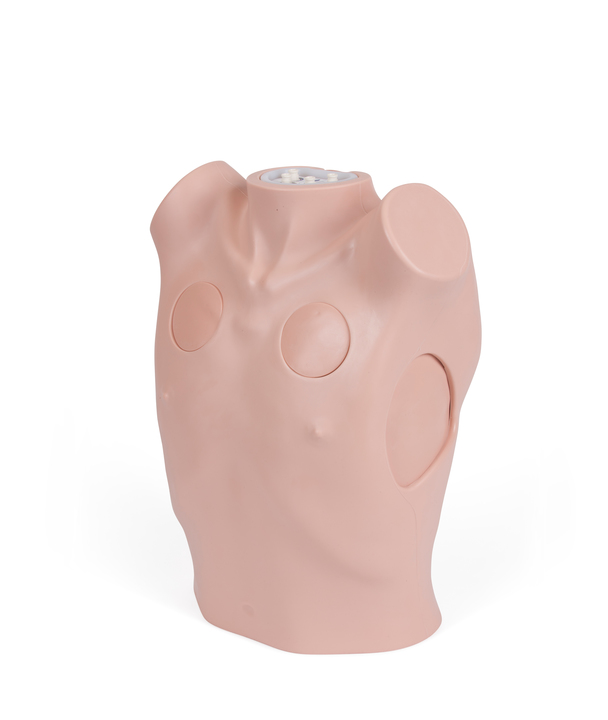
Developed to meet the training needs of healthcare professionals, this versatile simulator supports hands-on practice in surgical and guidewire-assisted thoracostomy, thoracentesis, chest drain insertion, and needle decompression.
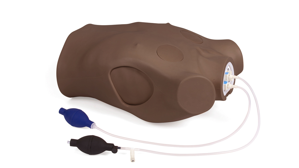
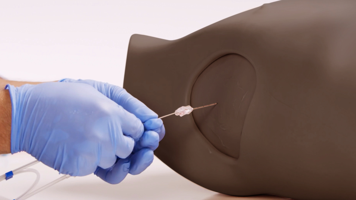
Both Standard and Advanced models allow chest tube insertion, with the Advanced version featuring echolucent material for ultrasound-guided Seldinger technique training.
Needle Decompression Pads
Positioned along the midclavicular lines, Needle Decompression Pads are designed to simulate the management of tension pneumothorax with realistic feedback—such as the audible release of air (“hiss”) or visible bubbles in a saline-filled syringe—upon successful chest cavity decompression.
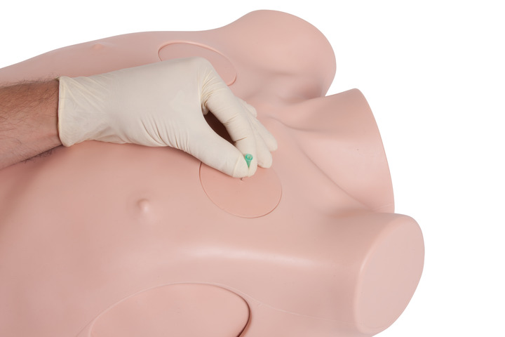
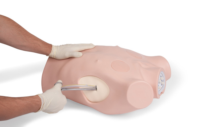
Standard Pads
Each Standard Pad features an integrated pleural layer that delivers a realistic “pop” sensation when a finger or forceps is inserted into the pleural space—mimicking the tactile feedback of real-life procedures. Constructed from high-quality foam, these pads are compatible with adhesive dressings and can be sutured to securely anchor a chest tube during training.
Advanced Pads
Designed for advanced simulation, these pads can be used with mock fluids to replicate pleural effusions or haemothorax during ultrasound-guided procedures. When connected to the included Respiratory Swing pump, the system mimics chest cavity movement, enhancing realism and improving ultrasound training accuracy.
Transform your training experience with integrated Augmented Reality (AR).
Our AR Training Mats and companion app merge real-world MRI and CT scan data with the expertise of medical illustrators and digital designers to vividly reveal the internal anatomy of our task trainers.
Using the free L&T ART app, learners can explore detailed anatomical overlays—including musculature, organs, vessels, and skeletal structures—directly on and around the trainer. The intuitive interface allows users to move around the model, toggle between layers, and view anatomical cross-sections in real time.
Students can also access step-by-step digital procedures within the AR environment, offering a clear visual guide to each technique and its impact on the patient’s anatomy.